Up until now, microscopy (magnifying a set image or object) has been limited. Conventional light microscopes that use an eyepiece lens to magnify what is being observed through the objective lens do not reveal “fine-scale details” of cells. Although there are high-resolution electron microscopes capable of enlarging the image of tissues, molecules, and other features at extensive magnifications, they are very expensive. But MIT and Harvard Medical School researchers have devised a better, yet still affordable, method for more accurate imaging.
In 2015, a group of researchers led by Edward Boyden, an associate professor of biological engineering and brain and cognitive sciences at MIT, developed a technique capable of expanding a tissue sample to 100 times its original volume before imaging it. In a recent study at MIT and Harvard, scientists used this technique as a more accurate and cheap solution to distinguish early-stage breast lesions with high or low risk of progressing to cancer.

The University Network (TUN) spoke with Boyden to gain insight on his microscopy technique and the recent study surrounding breast lesions.
“For more than 100 years, conventional light microscopes have been vital tools for pathology,” Boyden told TUN. “However, fine-scale details of cells cannot be seen with these scopes. Expansion allows researchers to see features with a conventional light microscope that ordinarily could be seen only with an expensive, high-resolution electron microscope. It also reveals additional molecular information that the electron microscope cannot provide. Since many diseases of concern in modern medicine start with very subtle changes, e.g., in cancers or autoimmune diseases or in aging, it is critical to be able to see fine-scale details of cells to diagnose disease early and accurately.”
The Study
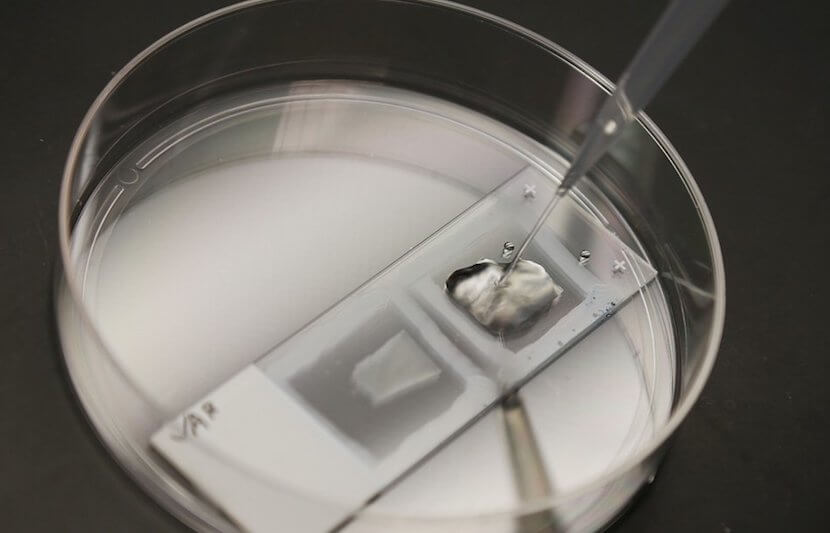
In the study, the researchers expanded the biopsy tissue samples. These tissue samples are commonly made more visible by being stained with a chemical, embedded in paraffin wax, or flash frozen.
To make the samples ready for expansion, the researchers exposed the samples to a chemical solvent called xylene to remove the chemical stain or paraffin. They then heated up the samples in another chemical named citrate.
Lastly, the samples go through a similar expansion process to the one developed in 2015. Boyden and his team embedded biological specimens in a dense swellable polymer and then added water. This made the biological specimen swell 100-fold in volume.
What differentiates the current study from the study in 2015 is that the researchers have to put the samples through stronger digestions steps because of the previous chemical fixation of the samples.
The researchers used this technique to test samples from patients with early stage breast lesions. A common way to test whether lesions will become malignant is by observing the nuclei of the cells. Lesions without typical nuclei have a much greater chance of progressing to cancer.
In the past, there have been many instances of misdiagnosis because of pathologists’ different assessments of atypical nuclei. A better way to differentiate benign lesions with atypical and typical nuclei could save hundreds of millions of dollars and prevent 400,000 yearly misdiagnosis in the U.S.
After expanding the tissue samples, the researchers used machine-learning algorithms to distinguish between lesions that were likely to become invasive and those that were not.
Additional Uses for Expansion
This approach can also be applied to find other diseases. In their analysis of kidney tissue, the researchers found that images of expanded samples showed signs of kidney disease that could previously only be found using an electron microscope.
“We analyzed kidney tissue samples from patients with nephrotic syndrome, which impairs the kidney’s ability to filter blood,” Boyden told TUN. “In these patients, tiny finger-like projections that filter the blood are lost or damaged. These structures are spaced about 200 nanometers apart and therefore can usually be seen only with an electron microscope or expensive super resolution microscopes.”
When the researchers showed the expanded tissue samples to a group of scientists, the group was able to identify the diseased tissue with 90 percent accuracy overall, compared to only 65 percent accuracy with unexpanded tissue samples.
Several times a month, Boyden and his team run training courses at MIT where visitors can attend and observe expansion microscopy techniques. Their goal is for many people to start using this approach to identify disease.
“Cancer biopsies are just the beginning,” Boyden said in a statement. “We have a new pipeline for taking clinical samples and expanding them, and we are finding that we can apply expansion to many different diseases. Expansion will enable computational pathology to take advantage of more information in a specimen than previously possible.”
Conclusion
Cancer and other autoimmune diseases are difficult to deal with physically, emotionally, and financially. A key factor in limiting the many hardships that accompany a disease is the ability to identify it early. However, many diseases, including cancer, start with very subtle changes that may not be caught early on. Approaches, such as this new expansion method, are therefore critical in the fight to quickly, cheaply, and accurately diagnose diseases.