
MIT researchers have created a smartphone-sized 3D ultrasound system designed to make breast cancer screening more frequent, affordable and accessible. The portable device could help catch aggressive tumors earlier, especially for people far from major hospitals.
For people at high risk of breast cancer, waiting a year between mammograms can feel like a lifetime. Tumors can grow in the gaps between annual screenings, and those so-called interval cancers are often more aggressive and harder to treat.
MIT researchers have now developed a portable, low-cost 3D ultrasound system that could make it much easier to scan breast tissue more often — not just in major hospitals, but in small clinics and, eventually, at home.
The miniaturized system pairs a small ultrasound probe, slightly smaller than a deck of cards, with a data processing module that is only a bit larger than a smartphone. When plugged into a laptop, it can reconstruct and display wide-angle 3D images of breast tissue in real time.
The compact design is central to the team’s goal of expanding access, noted senior author Canan Dagdeviren, an associate professor of media arts and sciences at MIT.
“Everything is more compact, and that can make it easier to be used in rural areas or for people who may have barriers to this kind of technology,” she said in a news release.
The work, published in the journal Advanced Healthcare Materials, was led by MIT doctoral candidate Colin Marcus and former postdoctoral researcher Md Osman Goni Nayeem, who collaborated with colleagues at MIT and Massachusetts General Hospital.
A push for more frequent screening
Mammograms, which use X-rays, remain the standard tool for breast cancer screening. But cancers that appear between yearly mammograms account for a significant share of cases and tend to be more aggressive than tumors found during routine scans.
When breast cancer is caught at its earliest stages, survival rates are extremely high. Once the disease is detected later, those odds drop sharply. That gap has driven interest in adding more frequent ultrasound scans for people at elevated risk, such as those with dense breast tissue or a strong family history.
Today, however, ultrasound is usually used only as a follow-up test if a mammogram flags something suspicious. The machines are large, expensive and typically housed in imaging suites at major hospitals and clinics. They also require trained technicians to operate.
“You need skilled ultrasound technicians to use those machines, which is a major obstacle to getting ultrasound access to rural communities, or to developing countries where there aren’t as many skilled radiologists,” added co-author Shrihari Viswanath, an MIT graduate student.
By shrinking and simplifying the technology, the MIT team hopes to make it feasible to scan more often — in community clinics, primary care offices and, one day, in people’s homes.
From bra-mounted patch to fully portable 3D
The new device builds on earlier work from Dagdeviren’s group. In 2023, her team created a flexible patch embedded with ultrasound transducers that could be attached to a bra. A separate tracker moved across the patch to capture 2D images from different angles, which could then be stitched together into a 3D view.
That first-generation system demonstrated the potential of wearable breast ultrasound, but it had key limitations. The images were generated by a traditional, refrigerator-sized ultrasound processing machine, and there could be small gaps in coverage between 2D slices, leaving room for tiny abnormalities to be missed.
In the new study, the researchers redesigned the hardware from the ground up to be fully portable and to capture true 3D images with fewer scan positions.
At the heart of the system is a chirped data acquisition system, or cDAQ, made up of the handheld probe and a custom motherboard. The probe’s ultrasound array is arranged in the shape of an empty square, a geometry that allows it to capture 3D information from the tissue beneath it.
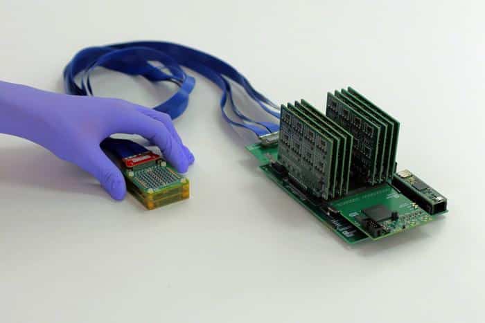
Caption: The new system consists of a small ultrasound probe, on left, attached to an acquisition and processing module that is a little larger than a smartphone.
Credit: Conformable Decoders Lab at the MIT Media Lab
The motherboard, built entirely from commercially available electronics, processes the incoming data. It is small enough to hold in one hand and costs about $300 to make, according to the team. When connected to a laptop, it can display 3D images of the breast in real time.
MIT Provost Anantha Chandrakasan, a co-author on the paper, emphasized how different this is from conventional systems.
“Traditional 3D ultrasound systems require power expensive and bulky electronics, which limits their use to high-end hospitals and clinics,” he said in the news release. “By redesigning the system to be ultra-sparse and energy-efficient, this powerful diagnostic tool can be moved out of the imaging suite and into a wearable form factor that is accessible for patients everywhere.”
Because the new device uses far less power than a standard ultrasound machine, it can run on a simple 5-volt DC supply — the same kind of power used for many small consumer electronics. That opens the door to battery-powered or plug-in use in settings that lack specialized infrastructure.
Reimagining ultrasound beyond the hospital
For decades, ultrasound imaging has been tightly linked to hospital environments and specialized equipment. The team set out to change that.
“Ultrasound imaging has long been confined to hospitals,” added Nayeem. “To move ultrasound beyond the hospital setting, we reengineered the entire architecture, introducing a new ultrasound fabrication process, to make the technology both scalable and practical.”
In early testing, the researchers used the system on a 71-year-old woman with a history of breast cysts. The device successfully imaged the cysts and produced a continuous 3D view of the surrounding tissue without gaps.
The probe can image up to 15 centimeters deep, and the team reports that scanning from just two or three positions is enough to cover the entire breast. Unlike traditional ultrasound exams, which often require the operator to press the probe firmly into the tissue, this device rests lightly on the skin.
That gentle contact helps preserve the accuracy of what clinicians see.
“With our technology, you simply place it gently on top of the tissue and it can visualize the cysts in their original location and with their original sizes,” Dagdeviren added.
What comes next
The researchers are now running a larger clinical trial at the MIT Center for Clinical and Translational Research and at Massachusetts General Hospital to further evaluate the device’s performance.
At the same time, they are working to shrink the electronics even more. Their goal is a data processing system about the size of a fingernail that could connect directly to a smartphone. That would eliminate the need for a separate module and laptop, making the entire setup smaller, lighter and easier to use.
The team also plans to develop a smartphone app powered by artificial intelligence to help guide users to the best locations to place the probe on the breast. That kind of guidance could be critical if the device is eventually used by patients at home rather than by trained clinicians.
While the current version could be adopted relatively quickly in doctors’ offices and imaging centers, the researchers ultimately envision a fully wearable sensor for people at high risk of breast cancer. Such a device could be integrated into clothing and used regularly to monitor for changes over time.
Dagdeviren is working to launch a startup to commercialize the technology, with support from MIT entrepreneurship and innovation programs and external funders. The research itself was backed by the National Science Foundation, industry partners and philanthropic foundations.
If the device continues to perform well in trials and makes its way into clinical practice, it could help shift breast cancer screening from a once-a-year event to a more continuous, personalized process — especially for those who need it most but have the least access to advanced imaging today.